


Lab 14: Blood and Blood Typing
Overview
The cardiovascular system includes three main components: the blood, blood vessels, and the heart. The heart serves as a pumping mechanism to distribute blood through the blood vessels and deliver nutrients, oxygen, and other important components to the tissues while removing waste and carbon dioxide. In order to operate effectively, all parts of the cardiovascular system must be functioning. In this lab, we will examine the anatomy of human blood using a microscope and explore how blood typing is performed.
Part 1: Blood Cells
Preliminary Information
Human blood is composed of plasma and cells. Plasma is the liquid part of the blood made up of water. Other components found in plasma include proteins, glucose, and hormones. There are three different types of blood cells: red blood cells (erythrocytes), white blood cells (leukocytes), and platelets (thrombocytes). Each of these has different shapes and functions.
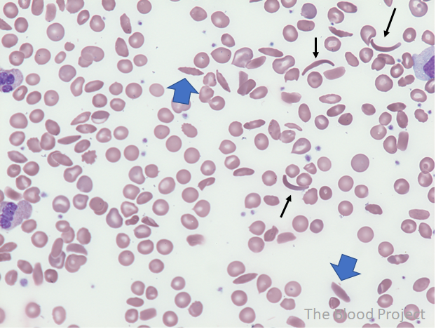
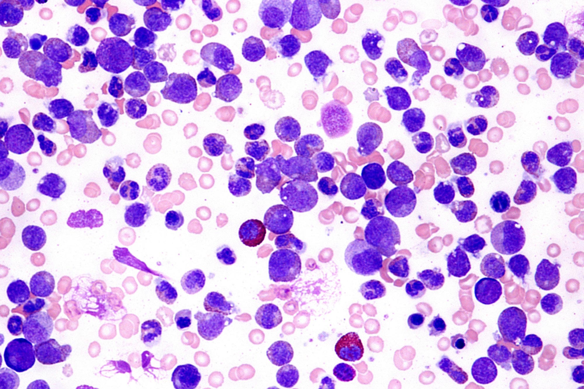
Some diseases can be diagnosed by looking at the appearance of blood cells or the relative number of blood cells. Sickle-cell anemia is a disease in which the erythrocytes become sickle or crescent shaped when oxygen levels are low. The crescent shaped cells can get caught in smaller blood vessels and, as a result, oxygen delivery is impaired. The term leukemia is a reference to a group of cancers that impacts the leukocytes. In a healthy individual, there are actually relatively low numbers of leukocytes in the blood compared to erythrocytes. Most leukocytes remain in other areas like the red bone marrow or other organs like lymph nodes or the spleen. With leukemia, there is an overproduction of abnormal leukocytes in the red bone marrow and the excess leukocytes end up moving into the blood. The abnormal leukocytes do not function normally and the overabundance of them in the bone marrow crowds out the normal blood stem cells causing various health complications, including anemia.
Lab Objectives
- Discuss the main components of blood and their importance in the human body.
- Differentiate between the blood of a healthy individual and that of someone with a blood disorder such as leukemia and sickle cell anemia.
Hypotheses:
Construct a hypothesis regarding the visible differences between the healthy blood cells and those on the sickle cell anemia slide:
|
|
Construct a hypothesis regarding the visible differences between the healthy blood cells and those on the infected blood slide:
|
|
Materials
- Reliable Internet
- Colored pencils
Methods
- Go to the website listed below and view the human blood smearthat represents a healthy human blood. Click on the image to make the image larger. Draw a small, but representative section of what you see in the area provided. Be sure to use colored pencils, markers or crayons.
https://imagebank.hematology.org/getimagebyid/3666?size=3
If for some reason this link does not work, Internet search prepared individual microscope slide human blood smear
- Label an erythrocyte, leukocyte, and thrombocyte (platelet) in your drawing.Erythrocytes will appear round and red. The main function of erythrocytes is to carry oxygen and iron. Erythrocytes are also the most abundant cells in the blood.
All leukocytes are round and purple with a nucleus. However, the size and shape of the nucleus varies. There are five different types of leukocytes found in the blood: neutrophils, monocytes, basophils, eosinophils, and lymphocytes. Some leukocytes contain granules in their cytoplasm which makes them appear grainy. The main function of the leukocytes is to protect the body from infections. They are the main players in our immune system.
Thrombocytes are cellular fragments and have no nucleus. They will appear as small purple specks/spots. The main function of thrombocytes is to stop bleeding by clotting the blood.
- Count the number of erythrocytes, leukocytes, and thrombocytes that you see in one field of view (one field of view is the entire area you see in the light). To count the erythrocytes, divide the field of view into quadrants (4 sections) and count the number you see in one quadrant and multiple by 4. Write your results in Table 1 within the Data section.
- Go to the website listed below and view the sickle-cells blood smearthat represents the human blood of a person with sickle cell trait or sickle cell disease. Draw a representative section of what you see in the area provided and be sure to include the sickle shaped red blood cell. Be sure to use colored pencils, markers or crayons.
https://www.thebloodproject.com/wp-content/uploads/2021/10/image-146.png
If for some reason this link does not work, Internet search Sickle Cell Anemia Blood Smear
- Label a SICKLED erythrocyte, a leukocyte, and a thrombocyte in your drawing.
- Countthe number of erythrocytes, leukocytes, and thrombocytes that you see in one field of view. Use the quadrant method explained in step 3 to estimate the number of erythrocytes. Write your results in Table 1 within the Data section.
- Note differences in the shapes and sizes of the blood cells compared to the healthy human blood slide. Emphasize these differences in your sketch.
- Go to the website listed below and view the of a leukemia blood smearthat represents the human blood of a person with an infection. Draw a representative section of what you see in the area provided. https://scitechdaily.com/images/Leukemia-Blood-Smear.jpg
If for some reason this link does not work, search for Leukemia Blood Smear
- Label at least 1 erythrocyte, leukocyte, and thrombocyte in your drawing.
- Countthe number of erythrocytes, leukocytes, and thrombocytes that you see in one field of view. Use the quadrant method explained in step 3 to estimate the number of erythrocytes. Write your results in Table 1 within the Data section.
- Note differences in the shapes and sizes of the blood cells compared to the healthy human blood slide. Emphasize these differences in your sketch.
Data
Table 1. Number of erythrocytes, leukocytes, and thrombocytes in microscope slides.
| Microscope Slide | Number of erythrocytes | Number of leukocytes | Number of thrombocytes |
| Human Blood Smear | |||
| Sickle-cell Anemia Blood | |||
| Leukemia Blood |
Conclusion – Discussion
- In the healthy blood smear, which cells were the most abundant?
|
|
- When compared to the erythrocytes in the healthy slide, what was different in the sickle cell anemia slide? How do you think that will affect the health of the individual affected?
|
|
- When compared to the leukocytes in the healthy slide, what was different in the leukemia blood slide? How do you think that will affect the health of the individual affected?
|
|
- Warfarin (aka Coumadin) is used as a blood thinner despite the fact that it does not thin the blood. Instead, it works to impair the process of clotting. What type of blood cells do you expect to be impacted by warfarin?
|
|
- Why would someone who has had a heart attack or stroke be prescribed warfarin? Explain your
answer.
|
|
Part 2: Blood Typing
Preliminary Information
All human cells contain markers called surface antigens built into the plasma membrane. These act as identification tags and allow the immune system to recognize the cells of an individual as ‘self’. For example, red blood cell membranes have surface antigens that determine blood type. Blood type is represented as a combination of the ABO blood antigen (Type A, B, AB, or O blood) and the Rh blood antigen (Rh+ (positive) when the Rh surface antigen is present, negative otherwise). For example, if a person has Type A+ blood, the red blood cells have the Type A surface antigen as well as the Rh antigen.
In order to protect the body, the immune system produces antibodies, proteins that attach to and attack ‘foreign’ antigens. Antibodies circulate in the blood and will cause a reaction if they encounter ‘non-self’ cell surface antigens. For example, if you are Type A+ blood, your immune system produces antibodies which will bind to and destroy any red blood cells that have the Type B surface antigens. These anti-B antibodies cannot bind to Type A surface antigens and do not attack red blood cells with Type A surface antigens. Table 2 outlines the major blood types of human blood.
Table 2. Blood Type, Surface Antigens, and Antibodies
| Blood Type | Red Blood Cell Surface Antigens | Antibodies Produced |
| A- | A | Anti-B
Anti-Rh |
| A+ | A, Rh factor | Anti-B |
| B – | B | Anti-A
Anti-Rh |
| B + | B, Rh factor | Anti-A |
| AB – | A and B | Anti-Rh |
| AB + (universal recipient) | A and B, Rh factor | None |
| O – (universal donor) | None | Anti-A
Anti-B Anti-Rh |
| O + | Rh factor | Anti-A
Anti-B |
Blood typing is particularly important from a medical perspective because there are many people who need donations of blood (blood transfusions) due to illness or injury. If an individual is given the wrong type of blood in a transfusion the immune system will attack the donated blood. This reaction by the immune system is called a transfusion reaction. The antibodies cause the donated red blood cells to clump and lyse (pop), which can lead to kidney failure and even death. When determining the types of blood that an individual can receive, it is important to focus on the red blood cells from the donated blood and to focus on the antibodies that the recipient possesses. You do not need to worry about the antibodies in the plasma from the blood of the donor because the antibodies are so diluted in the recipients blood that they do not usually cause an issue.
Lab Objectives
- Understand the importance of blood type in blood transfusions.
- Experience how blood typing is conducted.
Materials
- Watch the following Youtube videos:
- What are Blood Types?
- https://www.youtube.com/watch?v=ttjn1jVACk8
- Multiple Alleles (ABO Blood Types) and Punnett Squares
- https://www.youtube.com/watch?v=9O5JQqlngFY
Conclusion – Discussion
Complete the table below. One is done for you as an example.
| Person’s Blood Type | Types of Blood Individual Can Receive | Blood Type They Can Donate To |
| A+ | A+, A-, O+, O- | A+, AB+ |
| A- | ||
| B+ | ||
| B- | ||
| AB+ | ||
| AB- | ||
| O+ | ||
| O- |
- Why is blood type O- considered the universal donor? Be sure to use the vocabulary antigens and antibodies in your explanation, but do not copy from any Internet or other source.
|
|
- Why is blood type AB+ considered the universal recipient? Be sure to use the vocabulary antigens and antibodies in your explanation, but do not copy from any Internet or other source.
|
|
- Explain why it would be dangerous for someone who has a blood type of A- to receive a blood transfusion of AB- blood. Be specific. Be sure to use the vocabulary antigens and antibodies in your explanation, but do not copy from any Internet or other source.
|
|
- Monitoring the blood type of a pregnant female is very important because anti-Rh antibodies can cross over the placenta. Would a mother be concerned with this happening if she was Rh positive or negative? Explain your answer.
|
|
- The ABO blood system has 4 main blood types: A, B, AB, and O. Type A and B blood are both dominantly inherited, whereas, Type O is recessive. A couple has a baby and the man doesn’t think that the baby is his because the baby doesn’t look like him. His blood type is A and his partner’s blood type is O. The baby’s blood type is O and he is sure that proves this cannot be his baby. Using a Punnett Square, show the man whether or not this baby can be his or not.
| Possible Genotypes of the Offspring =
|
| Possible Phenotypes of the Offspring =
|
| Probability of these 2 parents having a child with O Blood Type =
|
- The ABO blood system has 4 main blood types: A, B, AB, and O. Type A and B blood are both dominantly inherited, whereas, Type O is recessive. Construct a Punnett square to predict the probability of having a child with Type O blood if your spouse has Type AB and you have Type O blood. Be sure to show the Punnett square, genotypes, and phenotypes. Once you’ve figured it out, write the probability of having a child with type O blood based on the given information.
| Possible Genotypes of the Offspring =
|
| Possible Phenotypes of the Offspring =
|
| Probability of these 2 parents having a child with O Blood Type =
|

